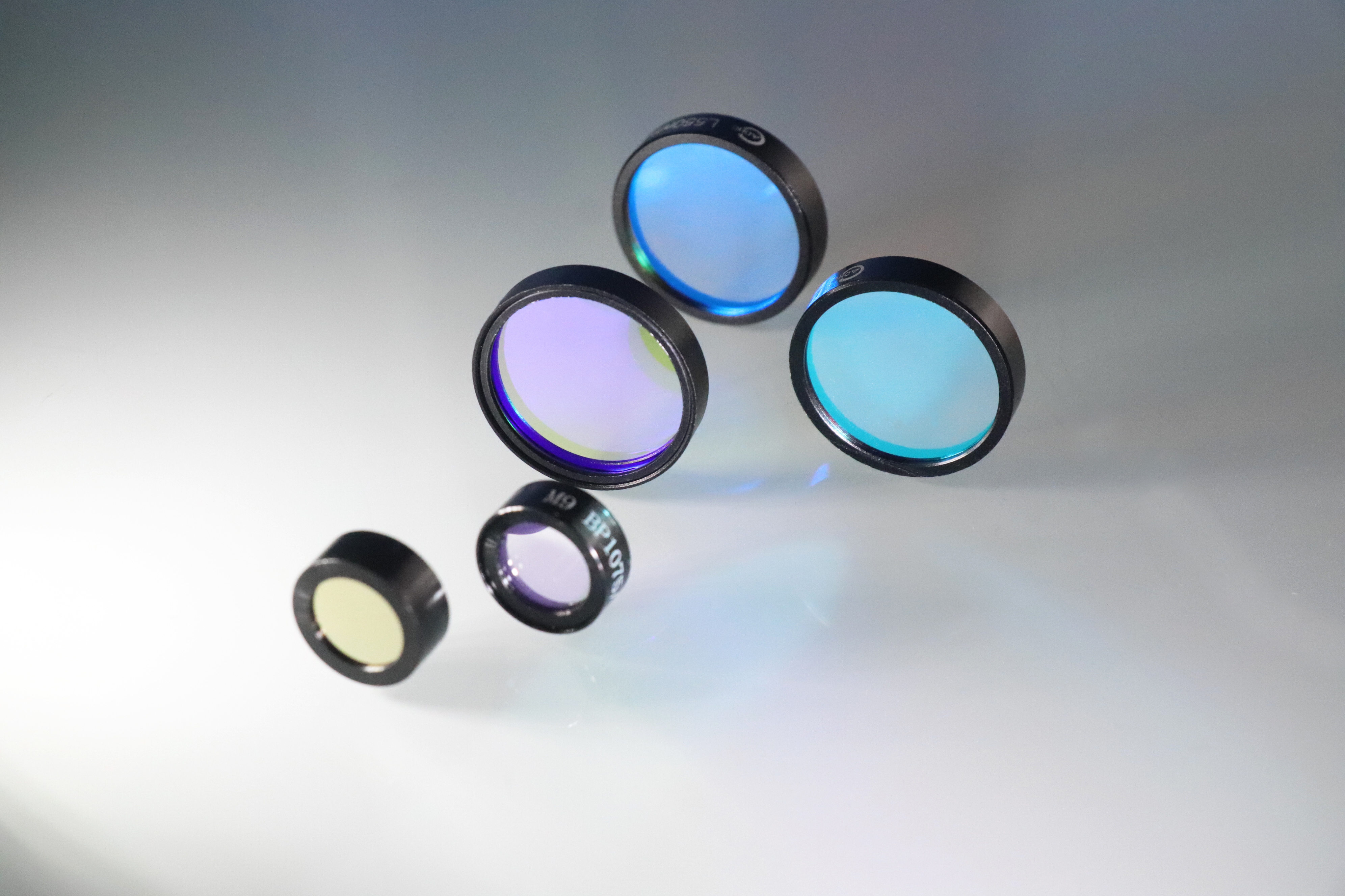
Fluorescence filters are optical devices typically made of special materials, used to manipulate the wavelength and intensity of light. Their primary function is to selectively transmit or block specific wavelengths of light, a property that makes them important in many fields.
The working principle of fluorescence filters is based on the absorption and transmission properties of light. Their design is usually based on the absorption and transmission spectra of specific materials. When light passes through a filter, only light of certain wavelengths can transmit through, while light of other wavelengths is absorbed or reflected by the filter, depending on the material properties.
Fluorescence is a process where a substance absorbs energy from an external light source, typically ultraviolet, blue, or other shorter wavelengths, causing electrons in the substance to transition from the ground state to an excited state. In the excitation process, the substance absorbs photons of light, raising the electrons to higher energy levels.
During the emission process, the excited electrons undergo non-radiative or radiative transitions back to the ground state. In this process, excess energy is released, often in the form of photons. These emitted photons typically have longer wavelengths than the excitation light source, resulting in fluorescence emission that is often part of the visible spectrum.
The specific wavelength of light emitted by fluorescence materials depends on their molecular structure and energy level distribution. Different fluorescent dyes or markers have distinct molecular structures, which determine their energy levels and fluorescence emission characteristics. When subjected to excitation light, only specific energy level transitions occur, leading to the emission of light at particular wavelengths.
Fluorescent materials are typically designed to emit light at specific wavelengths when excited by particular wavelengths of light. This design is based on the molecular structure and electronic energy levels of the fluorescent material. Through appropriate selection and design, fluorescent materials can achieve precise excitation and emission wavelengths, making them suitable for various fluorescence imaging and detection applications.
1. High Brightness Dyes:
Alexa Fluor: Alexa Fluor dyes are a series of high-performance fluorescent dyes known for their exceptional brightness and photostability. They are widely used in various fluorescence imaging and detection applications.
ATTO: ATTO dyes are a class of fluorescent dyes known for their high photostability and fluorescence quantum yield, suitable for a wide range of biological labeling applications.
DyLight: DyLight dyes are a class of fluorescent dyes characterized by their high fluorescence quantum yield and stability, suitable for various fluorescence labeling and imaging applications.
Qdot: Qdot dyes are semiconductor quantum dot fluorescent labels known for their narrow emission spectra and excellent brightness, used for high-sensitivity fluorescence imaging.
Quasar: Quasar dyes are fluorescent dyes used for nucleic acid probes and labeling, known for their excellent fluorescence quantum yield and photostability.
2. Organelle-specific Dyes:
LysoTracker: LysoTracker dyes are used to label lysosomes within cells, selectively entering lysosomes and emitting fluorescence signals.
MitoTracker: MitoTracker dyes are used to label mitochondria within cells, selectively entering mitochondria and emitting fluorescence signals.
3. Cellular Imaging Dyes:
FAM EX: FAM (Fluorescein Amidite) is a commonly used green fluorescent dye for nucleic acid labeling and staining.
Rhodamine: Rhodamine dyes are red fluorescent dyes used for cell labeling and fluorescence imaging.
Texas Red: Texas Red is a red fluorescent dye known for its high fluorescence quantum yield and stability, used in various fluorescence labeling and imaging applications.
4. Protein Labeling Dyes:
mCherry: mCherry is a red fluorescent protein used for live-cell imaging and protein labeling, commonly employed in cell tracking and localization.
5. pH-sensitive Dyes:
BCECF: BCECF (2',7'-Bis-(2-Carboxyethyl)-5-(and-6)-Carboxyfluorescein) is a pH-sensitive fluorescent dye used for measuring cellular pH changes, exhibiting dual fluorescence upon pH variation.
These dyes exhibit diverse fluorescence properties and are utilized in various biomedical and research applications, allowing for precise labeling, imaging, and analysis of biological specimens.
Figure 1. Bovine Pulmonary Artery Endothelial cell nuclei stained blue with DAPI, mitochondria stained red with MitoTracker Red CMXRos, and F-actin stained green with Alexa Fluor 488 phalloidin and imaged on a fluorescent microscope.
Different Types of Filters in Fluorescence Imaging
In fluorescence imaging and fluorescence microscopy, the filters commonly used can be categorized into the following three types based on their function and position:
1. Excitation Filters:
Excitation filters are positioned between the light source and the sample, selectively transmitting the excitation light wavelengths used to excite the fluorescent sample. They block the emitted fluorescence signal from the sample as well as other unnecessary light sources, thereby reducing background interference and improving imaging contrast and clarity.
2. Emission Filters (also known as Barrier Filters):
Emission filters are positioned between the sample and the detector, selectively transmitting the wavelength range of the fluorescent signal emitted by the sample. They block the excitation light and other non-relevant light sources, thereby eliminating background noise and enhancing imaging accuracy and sensitivity.
3. Dichroic Filters:
Dichroic filters are typically located in front of the detector and are used to adjust the relative intensity of different wavelength light and optimize imaging contrast and color balance. They can selectively transmit or block specific wavelength ranges of light as needed to adjust the optical path and optimize imaging.
These three types of filters play a crucial role in fluorescence imaging and microscopy, allowing for the acquisition of clear, high-contrast fluorescence images while excluding interference from non-relevant light sources.
Figure 2. Set up of fluorescence microscope.
Application of fluorescent filters
Fluorescence filters find extensive applications in fluorescence detection systems, including fluorescence microscopy, fluorescence microplate readers, fluorescence PCR analyzers, time-resolved fluorescence analyzers, and flow cytometers, among various medical analysis and detection instruments.
1. Fluorescence Microscopy:
Fluorescence microscopy stands as one of the most crucial tools in life science research. With light sources like mercury lamps, LEDs, or lasers, fluorescence filters are used to irradiate the specimen, causing it to emit fluorescence. Researchers then observe the shape and location of the specimen under a microscope. Fluorescence microscopy is employed in studying cellular processes such as absorption, transportation, and distribution of chemicals within cells.
2. Fluorescence Microplate Readers:
These readers utilize spectral scanning to determine the optimal excitation and emission wavelengths of unknown fluorescent dye molecules. They can detect a variety of fluorescent dye molecules, offering higher sensitivity levels for cellular experiments. Widely applied across organic chemistry, clinical diagnostics, drug screening, biochemistry, molecular biology, immunobiology, cell biology, environmental analysis, food safety testing, and material science.
3. Fluorescence PCR Analyzers:
These analyzers incorporate fluorescent dyes or probes into polymerase chain reactions (PCR), enabling real-time monitoring of the entire PCR process through the accumulation of fluorescence signals. They are extensively utilized in molecular diagnostics, molecular biology research, animal and plant quarantine, as well as food safety testing.
4. Time-Resolved Fluorescence Analyzers:
By delaying the measurement time according to the fluorescence spectrum of the fluorescent markers, these analyzers exclude non-specific fluorescence interference in the specimen, achieving precise quantitative analysis. They are commonly employed in the determination of proteins and peptide hormones, haptens, pathogen antigens, antibodies, tumor markers, dried blood spot samples, nucleic acids, and the activity of natural killer cells.
5. Flow Cytometers:
Flow cytometers are devices for automated analysis and sorting of cells. They rapidly measure, store, and display a range of important biophysical and biochemical characteristics of dispersed cells in a liquid medium. Additionally, they can select specific cell subsets based on predefined parameter ranges. Flow cytometry finds applications in immunology, oncology, hematology, and microbiology research, among others.
What does Shalom EO provide?
Shalom EO offers Fluorescence filters with a cutoff depth greater than OD6, making them ideal for various applications including Point-of-Care Testing (POCT), Polymerase Chain Reaction (PCR), and fluorescence microscopes. These filters are available in both stock and custom versions, ensuring flexibility to meet specific requirements. With industry-standard dimensions compatible with filter cubes from elite suppliers, Shalom EO filters offer seamless integration into existing systems. Moreover, they support a wide range of fluorophores, including FAM EX, Alexa Fluor, ATTO, BCECF, DyLight, LysoTracker, mCherry, MitoTracker, Qdot, Quasar, Rhodamine, Texas Red, and more, providing versatility and reliability for diverse fluorescence imaging and detection needs.


